40 drawing of microscope and label
How to draw compound of Microscope easily - step by step - YouTube Jul 6, 2019 ... I will show you " How to draw compound of microscope easily - step by step "Please watch carefully and try this okay.Thanks for watching. Microscope Imaging Software | Products | Leica Microsystems 20/08/2021 · A range of specific modules allows configuration of the microscope as a dedicated high-performance tool for almost any application. The latest software platform, LAS X, encompasses all microscope solutions for Life Science and Industry applications, offering maximum flexibility. The previous Leica Application Suite continues to be supported.
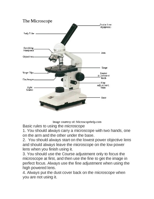
How to draw and label the parts of a microscope? What are at least ... The Eyepiece Lens. The Eyepiece Tube. The Microscope Arm. The Microscope Base. The Microscope Illuminator.Stage and Stage Clips. The Microscope Nosepiece. The ...

Drawing of microscope and label
Collection of Microscope Drawing (32) - Clipart Library Collection of Microscope Drawing (32). Satin Stitch Embroidery Design: Microscope Outline 3.50 inches H x. easy drawing of microscope. Label Microscope ... How To Draw A Microscope – A Step by Step Guide Microscope drawing in just 6 Easy Steps! · Step 1 · Step 2 – Now, draw the lenses for the microscope · Step 3 – Next, draw the arm and stage of the microscope ... Microscope Diagram Labeled, Unlabeled and Blank | Parts of a ... 1. Eyepiece/Ocular Lens – The lens into which the user looks to see the specimen. ... 3. Arm – A supporting piece of the optical microscope mounted upon the base.
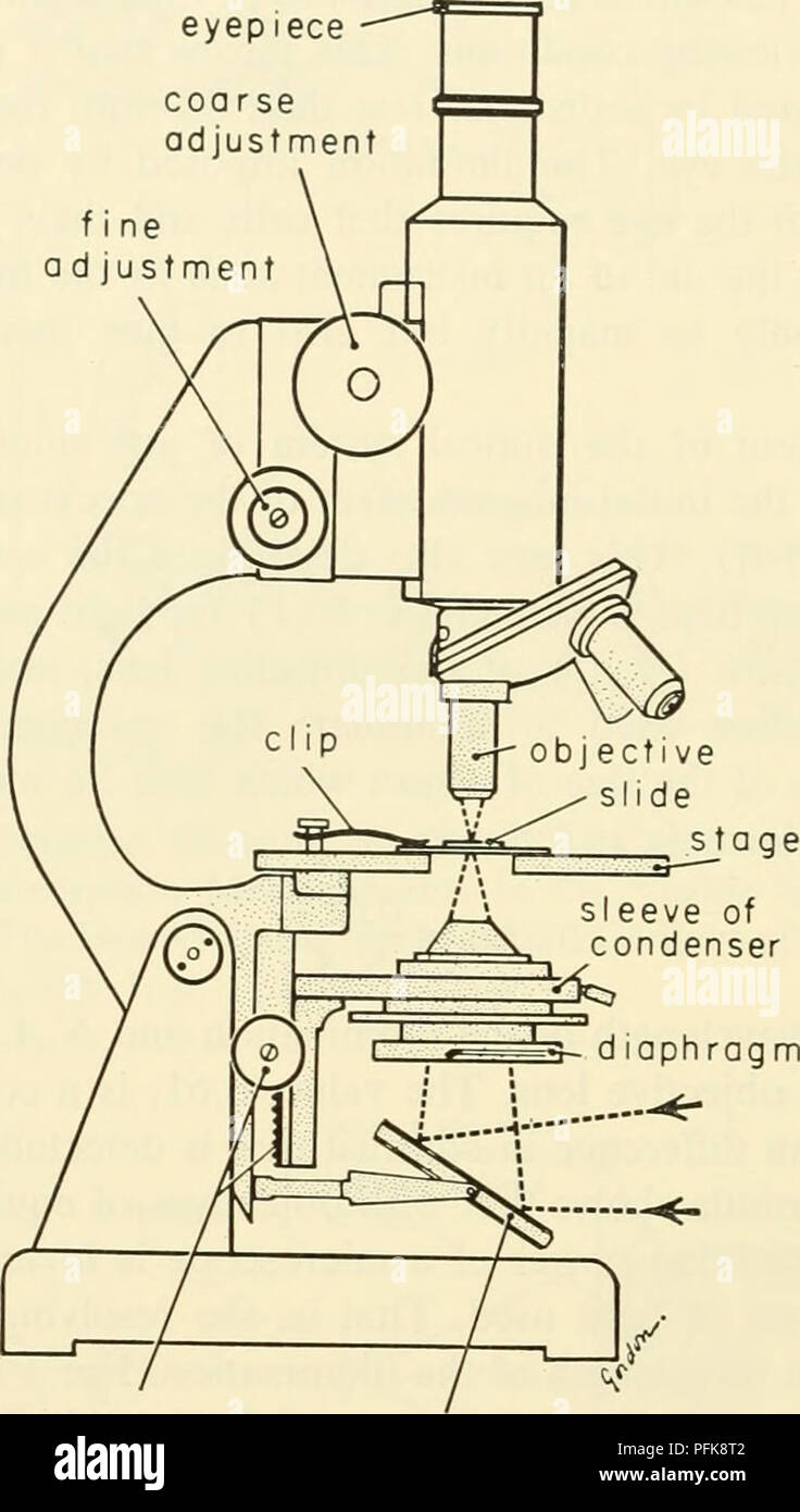
Drawing of microscope and label. Microscope Lab Experiments: An Introduction to the Microscope Microscope Worksheet: How to Record Microscope Observations In the field of science, recording observations while performing an experiment is one of the most useful tools available. Early scientists often kept very detailed journals of the experiments they performed, making entries for each individual experiment and writing down virtually everything they saw. Interactive Bacteria Cell Model - CELLS alive Periplasmic Space: This cellular compartment is found only in those bacteria that have both an outer membrane and plasma membrane (e.g. Gram negative bacteria).In the space are enzymes and other proteins that help digest and move nutrients into the cell. Cell Wall: Composed of peptidoglycan (polysaccharides + protein), the cell wall maintains the overall shape of a … A Study of the Microscope and its Functions With a Labeled Diagram 4 Best Microscope Drawing and Coloring Pages. This coloring page contains printable drawings various science lovers, including microscope instruments in ... Simple Microscope - Diagram (Parts labelled), Principle, Formula ... Feb 23, 2022 ... The working principle of a simple microscope is that when a lens is held close to the eye, a virtual, magnified and erect image of a specimen is ...
A new holographic microscope allows scientists to see through … 16/09/2022 · It is said that the new microscope can "see through" the intact skull, and is capable of high-resolution 3D imaging of the neural network within a living mouse brain without removing the skull. Cell Size and Scale - University of Utah Smaller cells are easily visible under a light microscope. It's even possible to make out structures within the cell, such as the nucleus, mitochondria and chloroplasts. Light microscopes use a system of lenses to magnify an image. The power of a light microscope is limited by the wavelength of visible light, which is about 500 nm. Microscope With Labels clip art - Pinterest Microscope Drawings handout. Description Use this blank handout as a way for students to record microscope drawings. Aside from the drawig itself, studnts are ... Microscope With Labels Clip Art at Clker.com how to draw a microscope easy · microscope drawing easy · compound microscope scatch · part of microscope and their functions · microscopi · parts of a ...
Lucy in the Sky with Diamonds - Wikipedia Legacy. Lennon mentioned "Lucy in the Sky" in the Beatles' song "I Am the Walrus".A 3.2-million-year-old, 40% complete fossil skeleton of an Australopithecus afarensis specimen, discovered in 1974 by Donald Johanson, Yves Coppens, Maurice Taieb and Tom Gray, was named "Lucy" because the Beatles song was being played loudly and repeatedly on a tape recorder in the camp. Microscope Lab Experiments: An Introduction to the Microscope Microscope Worksheet: How to Record Microscope Observations In the field of science, recording observations while performing an experiment is one of the most useful tools available. Early scientists often kept very detailed journals of the experiments they performed, making entries for each individual experiment and writing down virtually ... A new holographic microscope allows scientists to see through ... Sep 16, 2022 · It is said that the new microscope can "see through" the intact skull, and is capable of high-resolution 3D imaging of the neural network within a living mouse brain without removing the skull. Animal Cell Anatomy & Diagram - Enchanted Learning The cell is the basic unit of life. All organisms are made up of cells (or in some cases, a single cell). Most cells are very small; in fact, most are invisible without using a microscope. Cells are covered by a cell membrane and come in many different shapes. The contents of a cell are called the protoplasm. Glossary of Animal Cell Terms: Cell ...
How to draw microscope/Draw microscope in simple way.
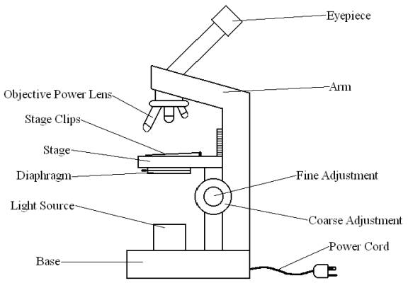
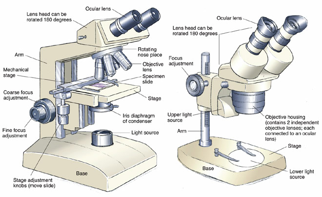
Parts of a microscope with functions and labeled diagram Sep 17, 2022 ... Head – This is also known as the body. It carries the optical parts in the upper part of the microscope. · Base – It acts as microscopes support.

Simple Microscope- Definition, Principle, Magnification ...
Microscope Imaging Software | Products | Leica Microsystems Aug 20, 2021 · A range of specific modules allows configuration of the microscope as a dedicated high-performance tool for almost any application. The latest software platform, LAS X, encompasses all microscope solutions for Life Science and Industry applications, offering maximum flexibility. The previous Leica Application Suite continues to be supported.

Clipart Panda - Free Clipart Images
Interactive Bacteria Cell Model - CELLS alive Ribosomes: Ribosomes give the cytoplasm of bacteria a granular appearance in electron micrographs.Though smaller than the ribosomes in eukaryotic cells, these inclusions have a similar function in translating the genetic message in messenger RNA into the production of peptide sequences (proteins).
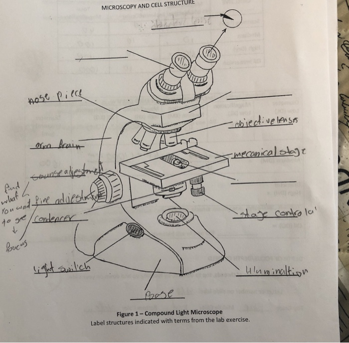
MICROSCOPY AND CELL STRUCTURE (C Figure 1- Compound | Chegg.com
Cell Size and Scale - University of Utah Smaller cells are easily visible under a light microscope. It's even possible to make out structures within the cell, such as the nucleus, mitochondria and chloroplasts. Light microscopes use a system of lenses to magnify an image. The power of a light microscope is limited by the wavelength of visible light, which is about 500 nm. The most powerful light microscopes can …

Draw a well labelled diagram of a microscope. - Brainly.in
Microscope Diagram Labeled, Unlabeled and Blank | Parts of a ... 1. Eyepiece/Ocular Lens – The lens into which the user looks to see the specimen. ... 3. Arm – A supporting piece of the optical microscope mounted upon the base.

How to Draw a Microscope and Label Its Parts
How To Draw A Microscope – A Step by Step Guide Microscope drawing in just 6 Easy Steps! · Step 1 · Step 2 – Now, draw the lenses for the microscope · Step 3 – Next, draw the arm and stage of the microscope ...
Free Microscope Drawing, Download Free Microscope Drawing png ...
Collection of Microscope Drawing (32) - Clipart Library Collection of Microscope Drawing (32). Satin Stitch Embroidery Design: Microscope Outline 3.50 inches H x. easy drawing of microscope. Label Microscope ...

How to draw microscope/Draw microscope in simple way. - YouTube
Cytology. Cytology. radiation used to illuminate the specimen ...
Free Microscope Drawing, Download Free Microscope Drawing png ...

How to draw Microscope diagram for beginners - step by step

How TO Draw microscope step by step easy/microscope drawing
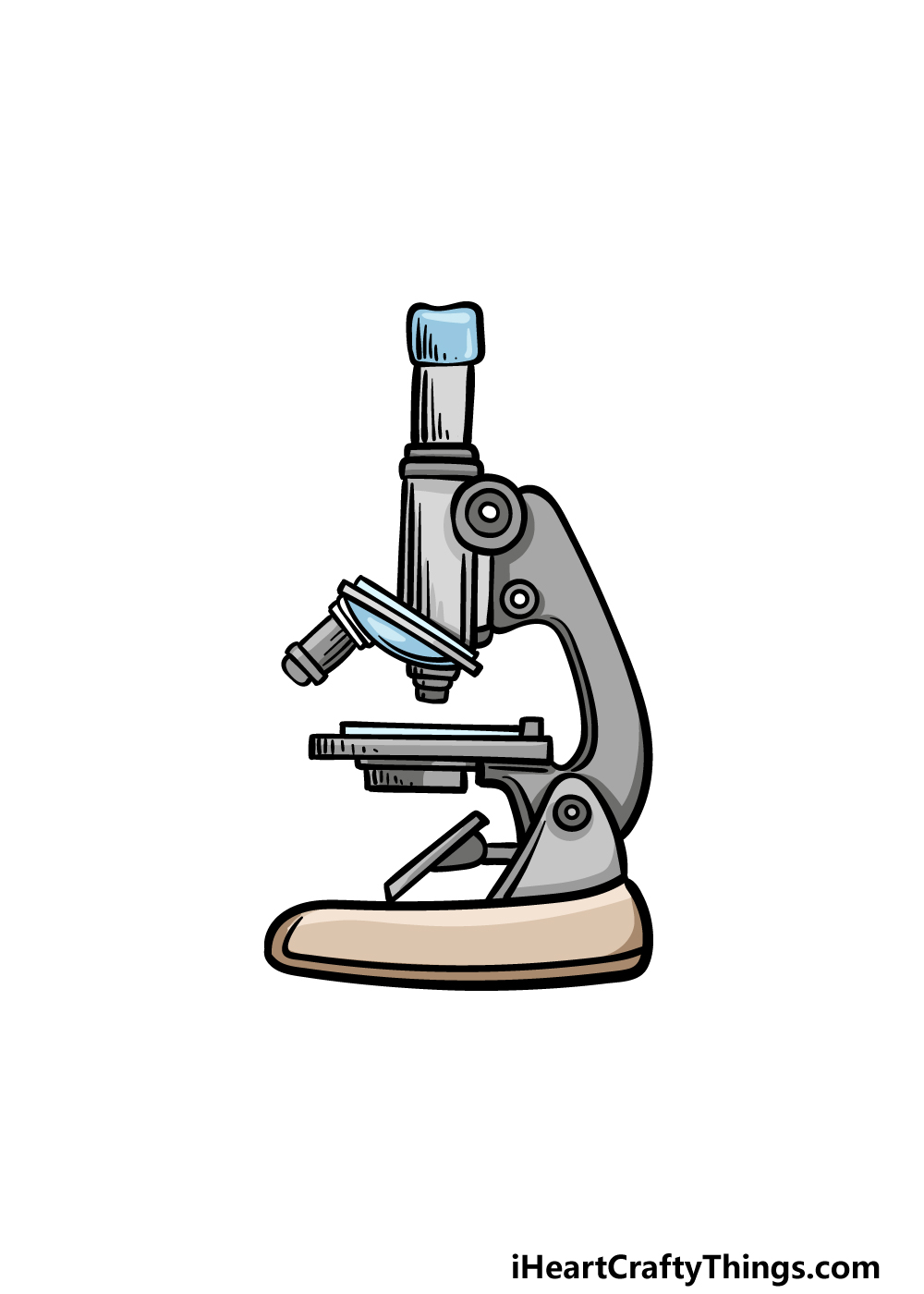
Microscope Drawing - How To Draw A Microscope Step By Step
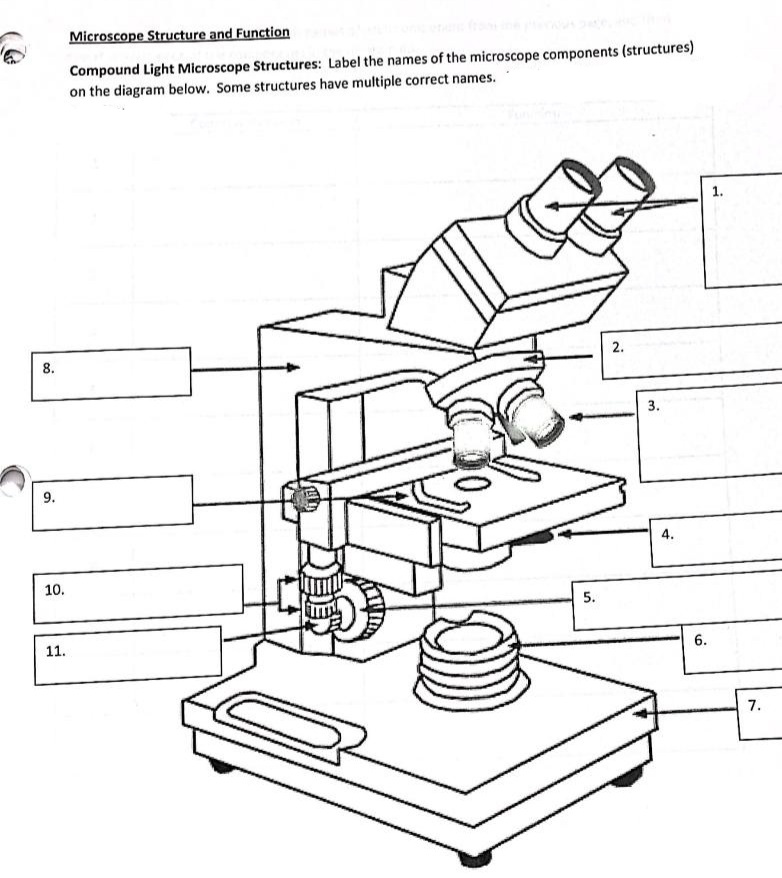
Answered: Microscope Structure and Function… | bartleby

Microscope - diagram Tom Butler | Science skills, Science ...
Free Microscope Drawing, Download Free Microscope Drawing png ...

Simple Microscope - Diagram (Parts labelled), Principle ...

Cell Drawing Microscope - Binocular Compound Microscope ...

How to Draw a Microscope Easy | Sketches easy, Easy drawings ...
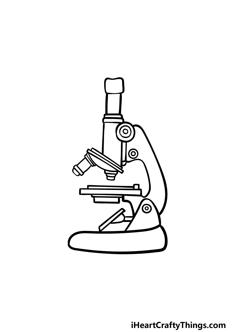
Microscope Drawing - How To Draw A Microscope Step By Step
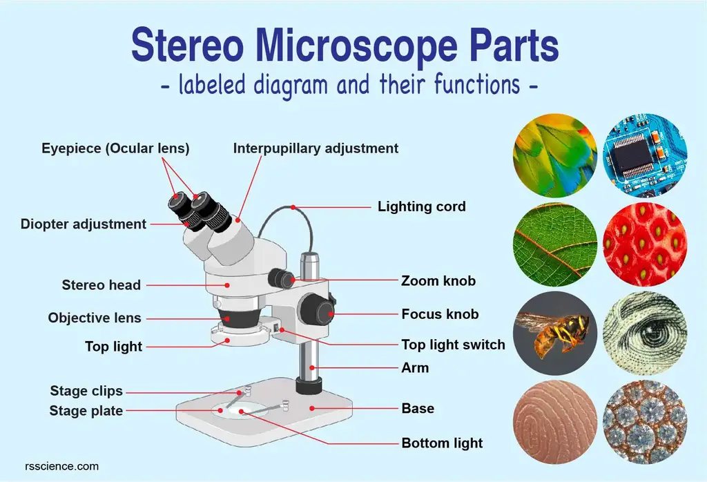
Parts of Stereo Microscope (Dissecting microscope) – labeled ...

Microscope Diagram Labeled, Unlabeled and Blank | Parts of a ...

Microscope Diagram Labeled, Unlabeled and Blank | Parts of a ...
Drawing microscope - Teaching resources

how to draw diagram of microscope | how to draw diagram of ...
Collection Of Free Microscopes Drawing Label Clipart ...

Label microscope pt.1 Diagram | Quizlet
Free Microscope Drawing, Download Free Microscope Drawing png ...

Compound microscope drawing | Clipart Panda - Free Clipart Images

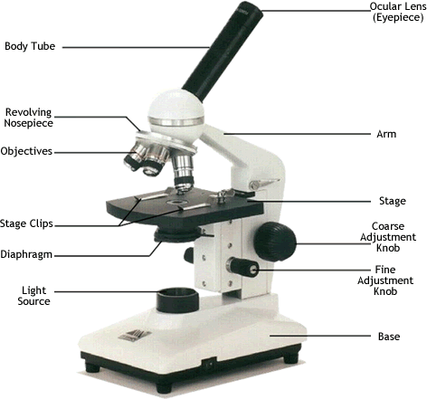
Compound Microscope Parts – Labeled Diagram and their ...

Microscope - Label - Part 2 Diagram | Quizlet
Parts of a microscope with functions and labeled diagram

Free Microscope Drawing, Download Free Microscope Drawing png ...

Simple Microscope - Diagram (Parts labelled), Principle ...

Labeling the Parts of the Microscope | Microscope World Resources

Parts of a microscope with functions and labeled diagram

Compound Microscope Parts – Labeled Diagram and their ...

Draw a labelled diagram of a compound microscope.

Compound Microscope Parts, Diagram Definition, Application ...

HOW TO DRAW MICROSCOPE


Komentar
Posting Komentar